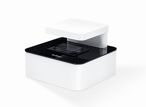
1. High-precision Automated Scanning Stage
The sample plate remains stationary while a built-in fully motorized XYZ 3D scanning stage moves to image all samples. This design eliminates cell displacement caused by plate movement.
2. Multiple Objectives & Multi-fluorescence Channels
Compatible with 4X/10X/20X/40X objectives, supporting overlay of brightfield and multi-channel fluorescent images.

3. Fluorescent Labeling & Multi-target Analysis
Specific fluorescent probes (fluorescent proteins/dyes) for distinct targets enable multi-target co-localization, interaction tracking, population & single-cell dynamic profiling, multi-parameter stress response monitoring, and transfection analysis.
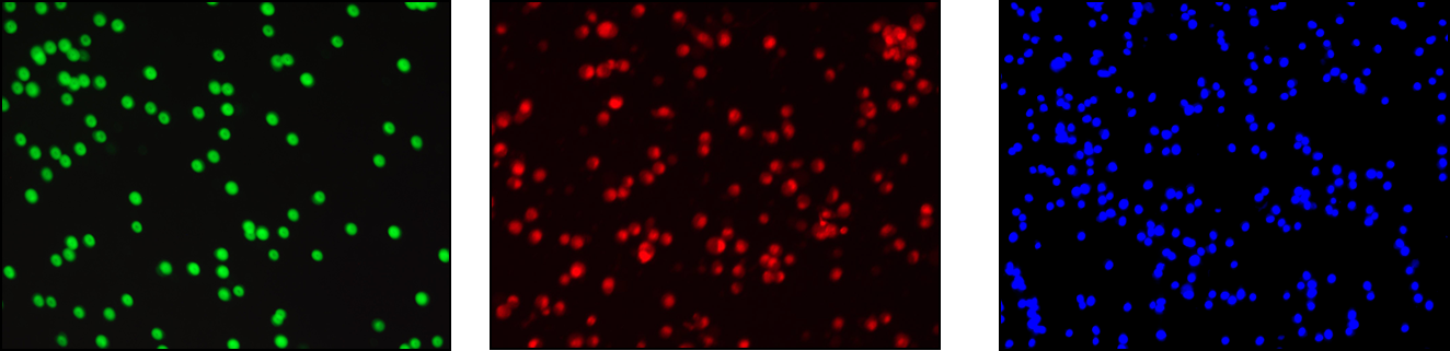
4. Image-based AI Data Analysis
AI identifies target cells via unique morphological features and outputs metrics including cell diameter, area, volume, confluency and fluorescence intensity. It supports comparative analysis across test/control groups with customizable screening parameters, and generates real-time charts and curves.

5. Live Cell Dynamic Imaging Analysis
The instrument fits inside CO₂ incubators for long-term live cell monitoring. Software automatically generates growth, confluency and migration curves per well, and exports dynamic imaging videos based on high-resolution cellular data.
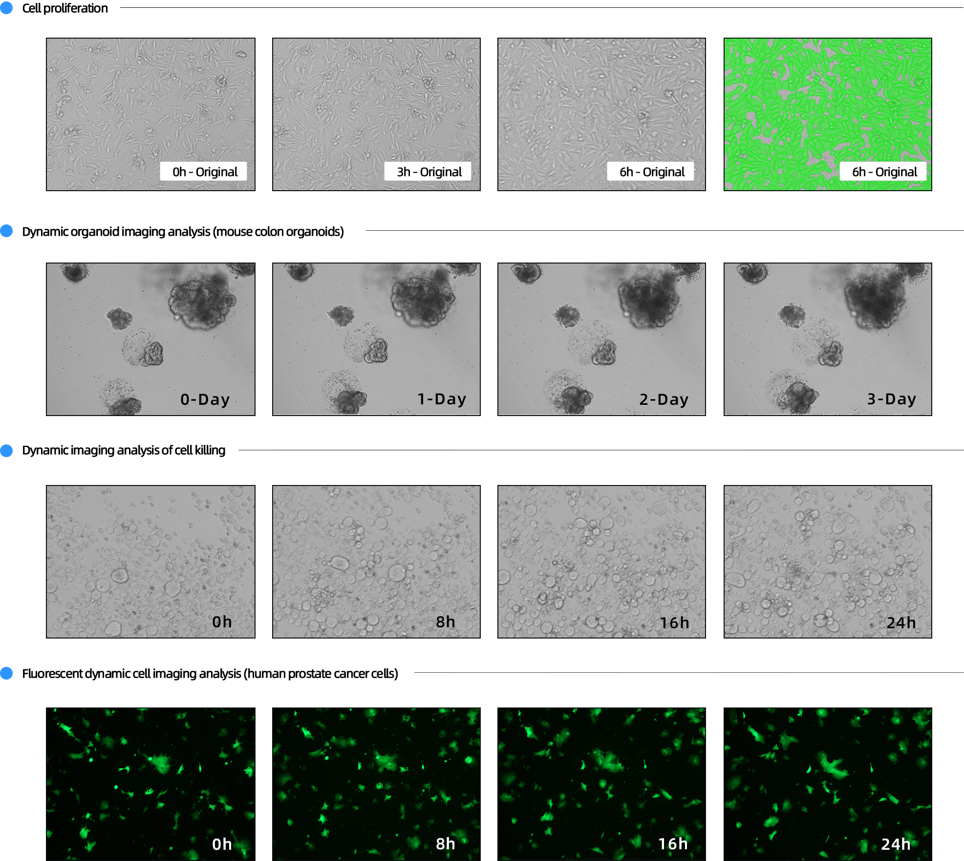
6. Z-axis Multi-layer Scanning
Micron-level Z-focus adjustment captures images at varying depths to acquire comprehensive morphological data (case: colorectal cancer organoids).

7. Compliant with FDA 21 CFR Part 11 & GMP Standards
8. Customizable Configuration


WeChat Official Account

Technical Support

