
Preface
Cell counting is an essential procedure in cell culture. Manual counting with a hemocytometer under a microscope is recognized as the gold-standard cell counting method. Here, Boda Boju introduces the detailed operating procedures of this gold-standard method for your reference.
* Prepare the Hemocytometer
1)Clean the surface of the hemocytometer and coverslip with absolute ethanol first.
2)Rinse the hemocytometer and coverslip with purified water afterwards.
3)Blow dry with a rubber pipette bulb after each cleaning.
4)Place the dedicated coverslip over the counting chambers of the hemocytometer.
Note: Avoid wiping the counting chambers of the hemocytometer to prevent damage to the engraved grid lines.
* Prepare Cell Suspension
1)For adherent cells, perform trypsinization to detach cells from the surface of culture flasks.
2)Vortex or repeatedly pipette the cell suspension to minimize cell clumping.
3)Suspension cells can be sampled directly for counting.
4)Optimal concentration of cell suspension for counting: \(5×10^5\) cells/mL – \(5×10^6\) cells/mL.
Notes:
a) Reduce cell clumps and keep clump size as small as possible.
b) If cell viability needs to be determined, mix the cell suspension with an equal volume of 0.4% trypan blue solution at a 1:1 volume ratio, mix well and incubate for 1–2 minutes.
* Load Cell Sample
1)Gently pipette the cell suspension to achieve uniform mixing.
2)Mix the cell suspension thoroughly with trypan blue solution at a volume ratio of 1:1.
3)Aspirate 10 μL of the cell suspension, and dispense the suspension onto the edge groove of the hemocytometer. The liquid will flow into the counting chamber under the coverslip via capillary action.
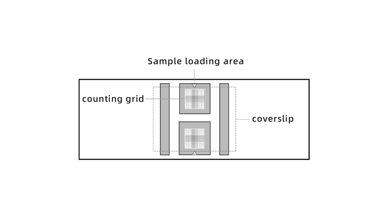
Notes:
Mix the cell suspension thoroughly before each loading to avoid increased sampling errors caused by cell sedimentation.
1)Load the cell suspension into the counting chamber in one continuous motion during sample loading to prevent bubble formation. The test must be repeated if bubbles appear.
2)Do not pipette the cell suspension directly into the counting chamber, as this will lead to uneven cell distribution.
4. Manual Cell Counting
Required equipment: Microscope equipped with a 10× objective lens, hemocytometer.
1)Allow the loaded hemocytometer to stand for several minutes after sample loading.
2)Place the hemocytometer on the microscope stage for observation, counting and data recording.
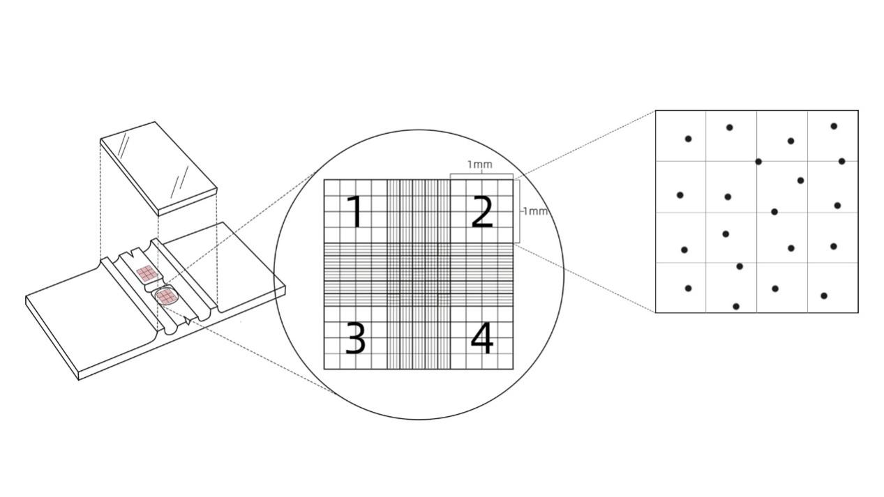
3)Count the total number of cells in four large squares numbered 1, 2, 3 and 4 respectively.
Notes:
1)It is recommended to repeat counting 1–2 times and calculate the average value to reduce counting errors.
2)The deviation of cell counts among the four regions should not exceed 5%. Reload the sample if the deviation exceeds the limit.
3)For cells crossing grid lines, follow the counting rule: count cells touching the top and left lines, exclude those touching the bottom and right lines.
4)To minimize counting errors, adjust the cell concentration to no less than 5×10⁵ cells/mL (more than 50 cells per large square).
5)Count only intact individual cells. Cell clumps should be counted as one single cell.
5. Calibration Standard for Hemocytometers
(Chinese Standard: JJG 552-1988 Trial Verification Regulation for Hemocytometers)
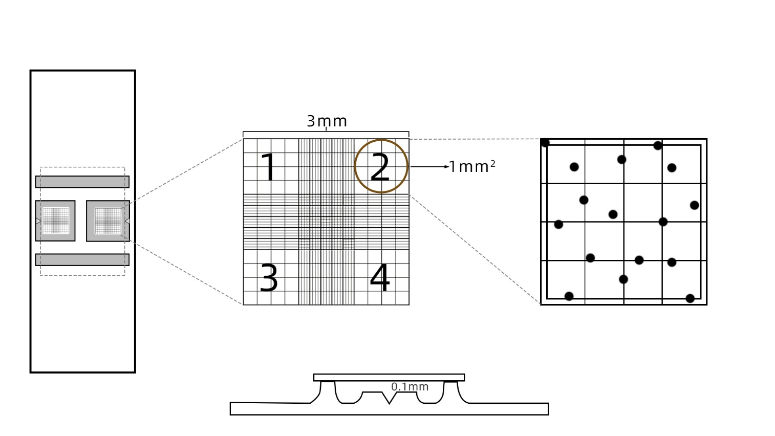
1)As shown in the figure above, after the coverslip is placed on the hemocytometer, the distance (depth) between the platform and the coverslip is 0.1 mm.
2)The central platform is precisely divided into 9 large squares, known as counting chambers. Each large square measures 3 mm × 3 mm with an area of 1 mm² and a volume of 0.1 mm³.
3)Cell concentration (cells/mL) = (Sum of cells counted in four large squares ÷ 4) × 2 (dilution factor for staining) × 10⁴.
Notes:
Do not multiply by 2 if trypan blue staining is not performed.
Many researchers may still encounter unsatisfactory results, low manual operation efficiency, unclear data readout, and eye strain from manual counting even after spending ample time learning and practicing the complete cell counting workflow.
Here we recommend an automated cell counter independently developed by Boda Boju, which supports standard hemocytometers!
6. Automated Cell Counter — Model JSY-SC-031N
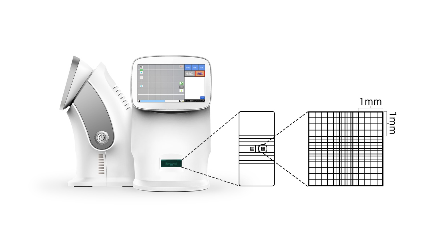
The Automated Cell Counter (Model JSY-SC-031N) is an integrated bright-field cell counter. It is equipped with a built-in 10× optical magnification system, automatic focusing unit, precision X-Y-Z three-axis linkage stage, and powerful cell recognition algorithm. The instrument is designed in accordance with the gold standard, namely the hemocytometer counting criteria.
Standard hemocytometers can be directly loaded onto the instrument. Manual rechecking under a microscope is available to avoid errors caused by repeated sample loading.
It automates and intelligently implements the gold-standard cell counting method, so you no longer need to doubt the accuracy of test results!
7. Real-scene Images of Loaded Hemocytometer on the Instrument
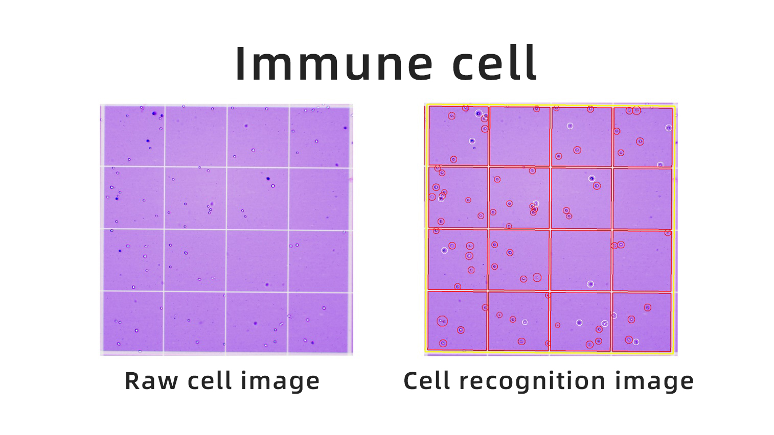
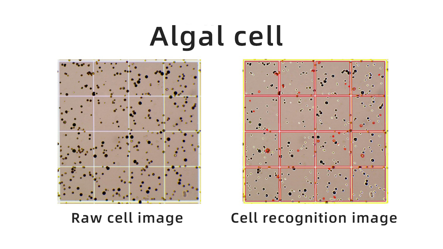
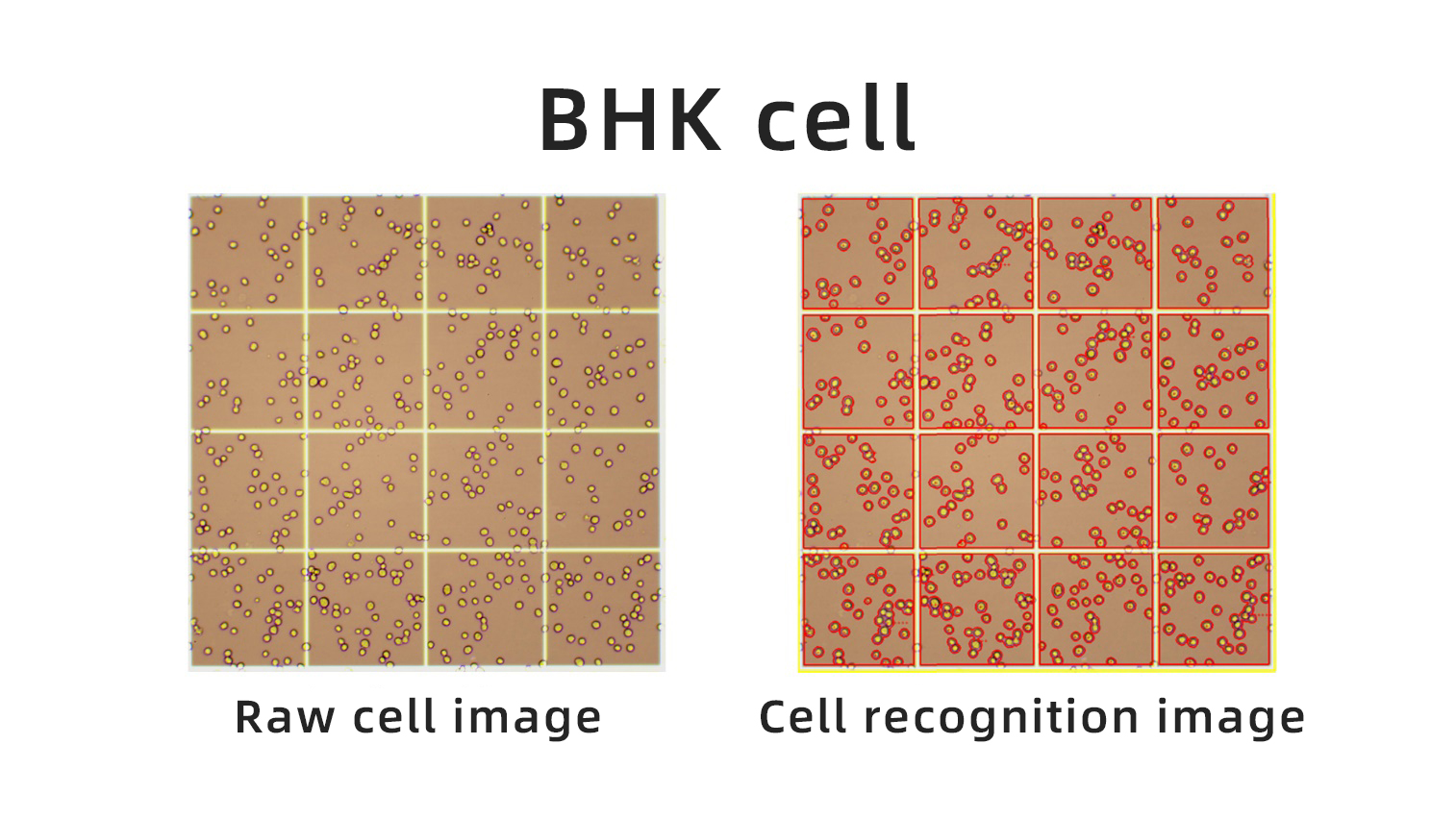
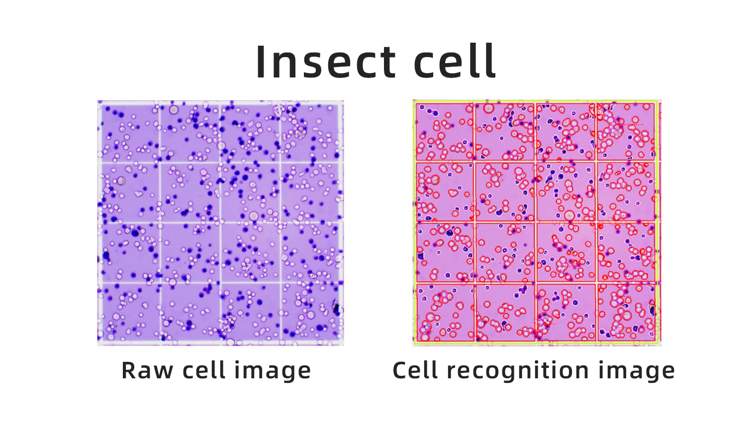 8. Cell Counting Results Include
Total cell concentration, viable/dead cell concentration, viable/dead cell ratio, total cell clump rate and concentration, viable cell clump rate and concentration, dead cell clump rate and concentration, raw images, recognized images, bar charts, scatter plots, contour plots, test reports, etc.
For more information, feel free to send us a message!


WeChat Official Account

Technical Support

